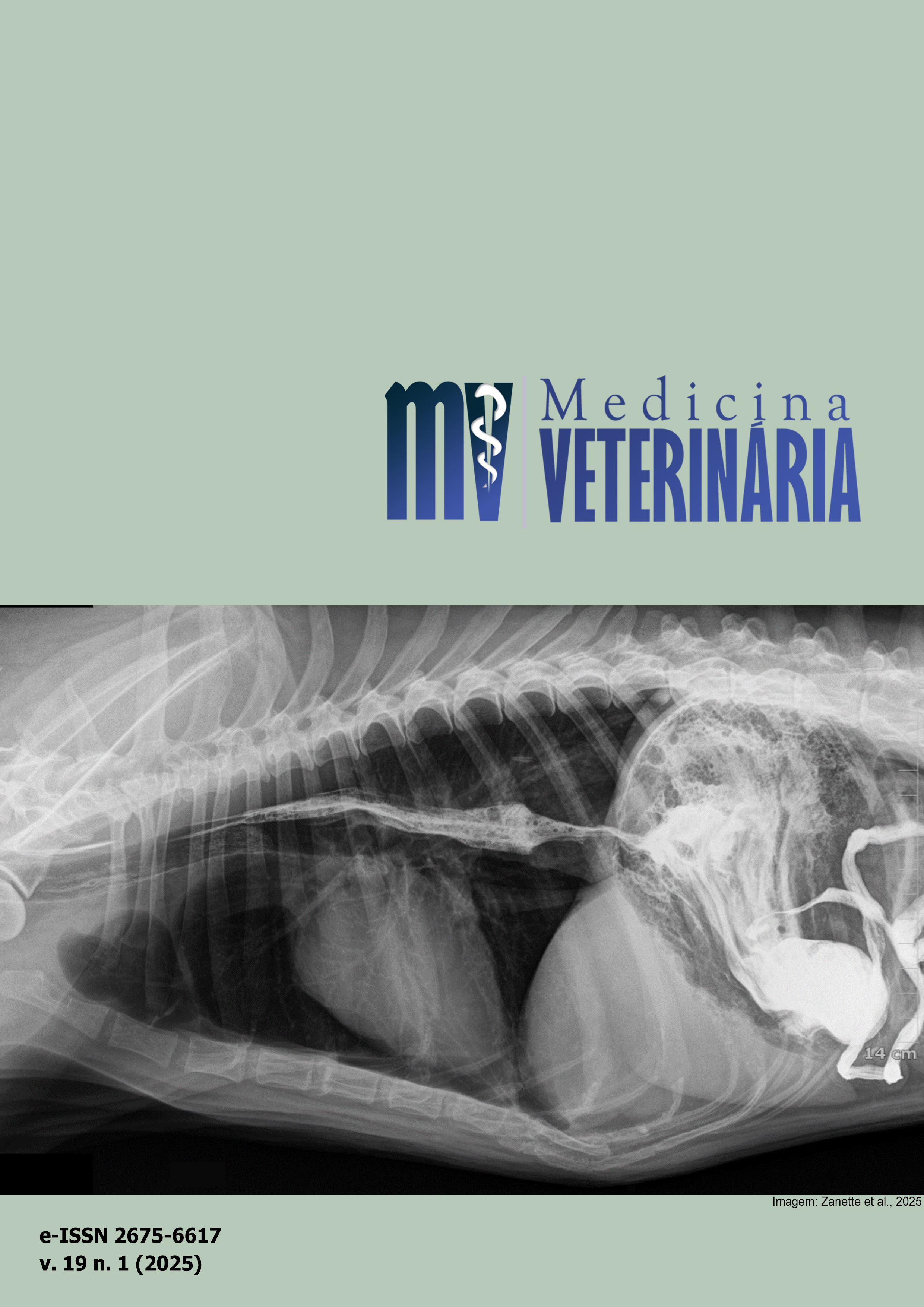
Detection of left atrial enlargement in dogs using deep learning in thoracic radiographs
DOI:
https://doi.org/10.26605/medvet-v19n1-6639Keywords:
computer-aided diagnosis, heart disease, image classifierAbstract
The study aimed to develop a tool to assist veterinarians in diagnosing left atrial enlargement on chest X-rays in dogs. The model utilized a total of 652 images all used in training and testing divided into two categories “positive” and “negative”. Three algorithms were used, obtaining the following results: the accuracy of the neural network was 89.7%, sensitivity of 90%, specificity of 89.5%, and Area Under the Curve (AUC) 95.8%. The accuracy of logistic regression was 88.2%, sensitivity 88.7%, specificity 87.8%, and AUC 94.1%. The decision tree accuracy was 69.6%, sensitivity 68.0%, specificity 71.0%, and AUC 69.6%. The classifier model with different algorithms can help radiologists improve the analysis of medical images by reducing errors, starting a selective double reading.Downloads
References
Abbott, J.A. Cardiomiopatia dilatada canina. In:_____. Segredos em cardiologia de pequenos animais. Porto Alegre: Artmed, 2006. p.304-314.
Alpaydin, E. Multilayer Perceptrons. In: Introduction to machine learning, 3rd ed. London: MIT Press, 2014. p.267-316.
Bahr, R.O Coração e os Vasos Pulmonares. In: Thrall, D.E. (Ed.). Diagnóstico de radiologia veterinária, 6a ed. Rio de Janeiro: Elsevier, 2015. p1220-1261.
Burti, S. et al. Use of deep learning to detect cardiomegaly on thoracic radiographs in dogs. The Veterinary Journal, 262: 1-7, 2020.
Derkatch, S. et al. Identification of vertebral fractures by convolutional neural networks to predict nonvertebral and hip fractures: a registry-based cohort study of dual x-ray absorptiometry. Radiology, 293(2): 1-7, 2019.
Du, D.Z.; Ko, K.I. Models of computation and complexity classes. In:_____. Theory of computational complexity. 2nd ed. Hoboken: John Wiley & Sons, 2011. p.3-44.
Godec, P. et al. Democratized image analytics by visual programming through integration of deep models and small-scale machine learning. Nature Communications, 10 (1): 1-7, 2019.
Gunderman, R.B.; Patel, P. Perception’s crucial role in radiology education. Academic Radiology, 26(1): 141–143. 2019.
Kealy, J.K.; McAllister, H. O tórax. In:_____. Radiologia e ultrassonografia do cão e do gato. 3a ed. Barueri: Manole, 2005. p.149-252.
Lamb, C.R. Veterinary diagnostic imaging: Probability, accuracy and impact. The Veterinary Journal, 215: 55-63, 2016.
Lamb, C.R.; Nelson, J.R. Diagnostic accuracy of tests based on radiologic measurements of dogs and cats: a systematic review. Veterinary Radiology & Ultrasound, 56(3): 231-244, 2015.
Li, S. et al. Pilot study: Application of artificial intelligence for detecting left atrial enlargement on canine thoracic radiographs. Veterinary Radiology & Ultrasound, 61(6): 611-618, 2020.
Liang, C.H. et al. Identifying pulmonary nodules or masses on chest radiography using deep learning: external validation and strategies to improve clinical practice. Clinical Radiology, 75(1): 38-45, 2019.
Malcolm, E.L. et al. Diagnostic value of vertebral left atrial size as determined from thoracic radiographs for assessment of left atrial size in dogs with myxomatous mitral valve disease. Journal of the American Veterinary Medical Association, 253(8): 1038-1045, 2018.
Rajpurkar, P. et al. Deep learning for chest radiograph diagnosis: A retrospective comparison of the CheXNeXt algorithm to practicing radiologists. PLoS Med, 15(11): 1-17, 2018.
Shorten, C.; Khoshgoftaar, T.M. A survey on Image Data Augmentation for Deep Learning. Journal of Big Data, 60(1): 1-48, 2019.
Thian, Y.L. et al. Convolutional Neural Networks for Automated Fracture Detection and Localization on Wrist Radiographs. Radiology: Artificial Intelligence, 1(1): 1-8, 2019.
Thrall, D.E. Introdução à Interpretação Radiográfica. In:_____. Diagnóstico de radiologia veterinária. 6a ed. Rio de Janeiro: Elsevier, 2015. p.175-209.
Ucar, F.; Korkmaz, D. COVIDiagnosis-Net: Deep Bayes-SqueezeNet based diagnosis of the coronavirus disease 2019 (COVID-19) from X-ray images. Medical Hypotheses, 140: 1-12, 2020.
Van Vleet, J.F.; Ferrans, V.J. Patologia do sistema cardiovascular. In: Carlton, W.W., McGavian, M.D., (Ed.). Patologia veterinária especial. 2a ed. Rio Grande do Sul: Artmed, 1998. p.194-227.
Webb, S. Technology feature deep learning for biology. Nature, 554(7693): 555-557, 2018.
White, S.C. Cysts of the jaws. In: White, S.C.; Pharoah, M.J. Oral Radiology: principles and interpretation. 5th ed. St. Louis: Mosby, 2004. p.355-377.
Downloads
Published
How to Cite
Issue
Section
License
Copyright (c) 2025 Lucas Auto Lopes, Anaemilia das Neves Diniz, Helio Silva Neto

This work is licensed under a Creative Commons Attribution-NonCommercial-ShareAlike 4.0 International License.
A Revista de Medicina Veterinária permite que o autor retenha os direitos de publicação sem restrições, utilizando para tal a licença Creative Commons CC BY-NC-SA 4.0.
De acordo com os termos seguintes:
Atribuição — Você deve dar o crédito apropriado, prover um link para a licença e indicar se mudanças foram feitas. Você deve fazê-lo em qualquer circunstância razoável, mas de nenhuma maneira que sugira que o licenciante apoia você ou o seu uso.
NãoComercial — Você não pode usar o material para fins comerciais.
CompartilhaIgual — Se você remixar, transformar, ou criar a partir do material, tem de distribuir as suas contribuições sob a mesma licença que o original.
Sem restrições adicionais — Você não pode aplicar termos jurídicos ou medidas de caráter tecnológico que restrinjam legalmente outros de fazerem algo que a licença permita.


