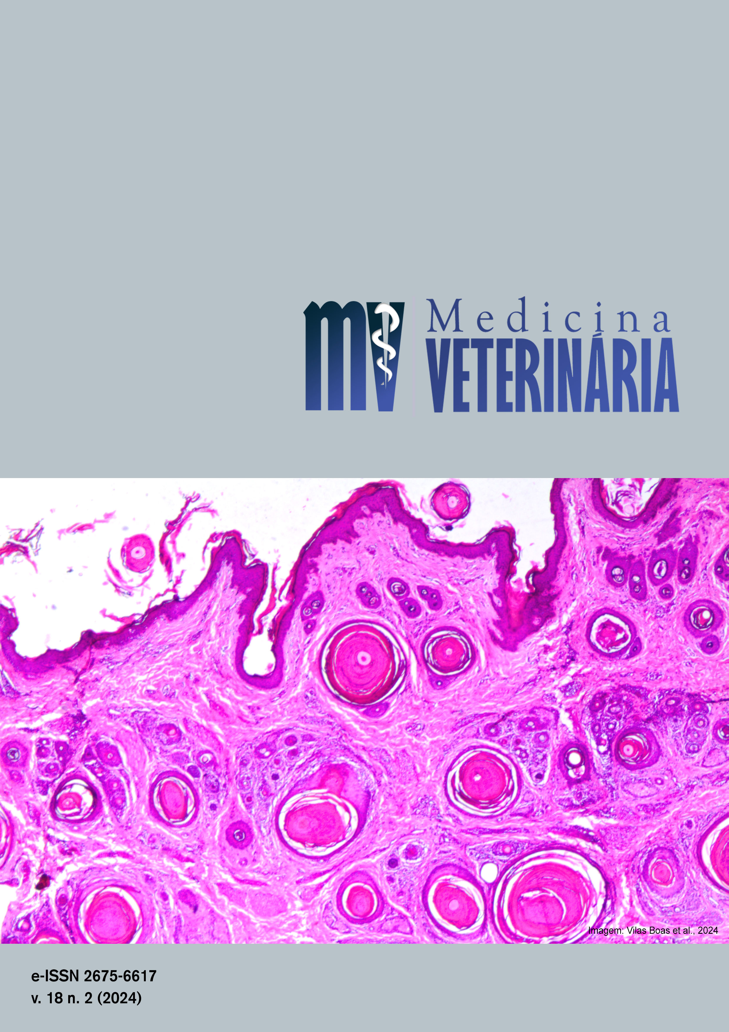
Perosomus acaudatus associado à atresia anal em bezerro: relato de caso
DOI:
https://doi.org/10.26605/medvet-v18n2-6672Keywords:
cattle, colostomy, malformation, peritonitisAbstract
This report aims to address the case of a calf presenting with Perosomus acaudatus associated with anal atresia. According to the owner, during the initial handling of the calf, observed the absence of the tail and anus. Subsequently, employees on the property performed a perforation in the perineal region. Upon being received by the Ruminant Clinic team at HVU-UFSM, the absence of the tail and anus was confirmed, with the presence of a healing wound in the perineal region and a longitudinal depression in the dorsal region of the pelvis. A radiographic examination revealed three non-fused sacral vertebrae and the absence of coccygeal vertebrae. Additionally, the colon and rectum were filled with gas. The owner was proposed to undergo surgical treatment for the correction of anal atresia. Due to adhesions of the rectum in the pelvic region, it was im possible to perform anoplasty. Instead, a colostomy was performed on the abdominal wall in the left inguinal region. After the procedure, the patient was able to defecate; however, in the following week, it exhibited apathy and prostration. A sample of peritoneal fluid was collected, revealing a change in color, turbidity, and an increase in nucleated cells. Given the findings, euthanasia was performed, followed by a necropsy of the animal. The observations included the absence of coccygeal vertebrae, the presence of only three sacral vertebrae, diffuse fibrinonecrosupurative peritonitis, diffuse transmural necrotizing colitis, and multifocal necrotizing dermatitis. The occurrence of Perosomus acaudatus, despite being rare, should always be closely examined to identify possible associated abnormalities. The outcome of this patient does not change the recommendation for surgical correction in cases of anal atresia.Downloads
References
Afonso, J.C.D.A.S.; Dantas, A.C.; Costa, N.A.; Mendonça, C.L.; Silva Filho, A.P. Achados clínicos de bovinos com úlcera de abomaso. Veterinária e Zootecnia, 19(2): 196-206, 2012.
Antonioli, M.L.; Carvalho, J.R.G.; Bustamante, C.C.; Mendonça, L.F.; Bergamasco, P.L.F.; Escobar, A.; Marques, L.C.; Canola, P.A. Anal atresia with rectovaginal fistula in sheep: case report. Arquivo Brasileiro de Medicina Veterinária e Zootecnia, 69(5): 1167-1171, 2017.
Arthur, G.H.; Noakes, D.E.; Pearson, H.; Parkinson, T.J. Types of dystocia within species. In: Noakes, D.E.; Parkinson, T.J.; England, G.C.W.; Arthur, G.H. (Eds). Arthur’s Veterinary reproduction and obstetrics. 8th ed. Philadelphia: W.B. Saunders, 2001. p.212-218.
Azizi, S.; Mohammadi, R.; Mohammadpour, I. Surgical repair and management of congenital intestinal atresia in 68 calves. Veterinary Surgery, 39(1): 115-120, 2010.
Clinical Pathology Laboratory, Cornell University. Hematology (Advia 2120), 2024. Disponível em: <https://www.vet.cornell.edu/animal-health-diagnostic-center/laboratories/clinical-pathology/reference-intervals/hematology>. Acesso em: 20 jun 2024.
Hendrickson, D.A. Técnicas cirúrgicas em grandes animais. 3a ed. Rio de Janeiro: Guanabara Koogan, 2007. 312p.
Hossain, M.B.; Hashim, M.A.; Hossain, M.A.; Sabrin, M.S. Prevalence of Atresia ani in new born calves and their surgical management. Bangladesh Journal of Veterinary Medicine, 12(1): 41-45, 2014.
Huston, K.; Wearden, S. Congenital taillessness in cattle. Journal of Dairy Science, 41(10): 1359-1370, 1958.
Jones, S.L.; Smith, B.P. Diseases of the Alimentary Tract. In: Smith, B.P. Large animal internal medicine. 8th ed. Missouri: Elsevier, 2013. p. 638-842.
Luna, S.P.L.; Carregaro, A.B. Anestesia e analgesia em equídeos, ruminantes e suínos. 1a ed. São Paulo: Medvet, 2019. 398p.
Noh, D.H.; Jeong, W.I.; Lee, C.S.; Jung, C.Y.; Chung, J.Y.; Jee, Y.H.; Do, S.H.; An, M.Y.; Kwon, O.D.; Williams, B.H.; Jeong, K.S. Multiple congenital malformation in a Holstein calf. Journal of Comparative Pathology, 129(1): 313-315, 2003.
Radostits, O.M.; Gay, C.C.; Blood, D.C.; Hinchcliff, K.W. Doenças dos recém-nascidos. In: Radostits, O.M.; Blood, D.C.; Gay, C.C.; Hinchcliff, K.W. Clínica veterinária, um tratado de doenças dos bovinos, caprinos e equinos. 9a ed. Rio de Janeiro: Guanabara Koogan, 2000. p.102-137.
Remi-Adewunmi, B.D.; Fale, M.S.; Usman, B.; Lawal, M. A retrospective study of atresia ani cases at the Ahamdu Bello University veterinary teaching hospital Zaria, Nigeria. Nigerian Veterinary Journal, 28(1): 48-53, 2007.
Roberts, S.J. Veterinary obstetrics and genital diseases. 2nd ed. India: CBS Publishers and Distributors, 1971. 776p.
Satheshkumar, S.; Punniamurthy, N. Perosomus acaudatus in a heifer. Indian Veterinary Journal, 84(10): 1174-1174, 2004.
Shivaprakash, B.V.; Ramakrishna, V.; Rao, D.G. Perosomus acaudatus, pelvic malformation and atresia ani et recti in a deoni calf. Indian Veterinary Journal, 72(4) 387-389, 1995.
Silva Filho, A.P.; Afonso, J.A.B.; Souza, J.C.A.; Dantas, A.C.; Costa, N.A.; Mendonça, C.L. Achados clínicos de bovinos com úlcera de abomaso. Veterinária e Zootecnia, 19(2): 196-206, 2012.
Teixeira, U.R.; Araújo, K.C. Atresia anal em bovino: um relato de caso. Revista Ibero-Americana de Humanidades, Ciências e Educação, 8(10): 1379-1390, 2002.
Yanaka, R.; Ferreira, H.N.; Assis, M.M.Q.; Oliveira, G.K.; Albuquerque, V.B.; Sartori, V.C. Multiple congenital malformations of a nellore calf: case report. Ars Veterinaria, 28(3): 144-147, 2012.?
Downloads
Published
How to Cite
Issue
Section
License
Copyright (c) 2024 Rodrigo Santos Severo de Souza, Henrique Ravalha e Siqueira, Caroline Garlet Dallanôra, Romario Stroeher, Rodrigo Dalmina Rech, Marcelo da Silva Cecim, Otavio Luiz Fidelis Junior, Marta Lizandra do Rêgo Leal

This work is licensed under a Creative Commons Attribution-NonCommercial-ShareAlike 4.0 International License.
A Revista de Medicina Veterinária permite que o autor retenha os direitos de publicação sem restrições, utilizando para tal a licença Creative Commons CC BY-NC-SA 4.0.
De acordo com os termos seguintes:
Atribuição — Você deve dar o crédito apropriado, prover um link para a licença e indicar se mudanças foram feitas. Você deve fazê-lo em qualquer circunstância razoável, mas de nenhuma maneira que sugira que o licenciante apoia você ou o seu uso.
NãoComercial — Você não pode usar o material para fins comerciais.
CompartilhaIgual — Se você remixar, transformar, ou criar a partir do material, tem de distribuir as suas contribuições sob a mesma licença que o original.
Sem restrições adicionais — Você não pode aplicar termos jurídicos ou medidas de caráter tecnológico que restrinjam legalmente outros de fazerem algo que a licença permita.


